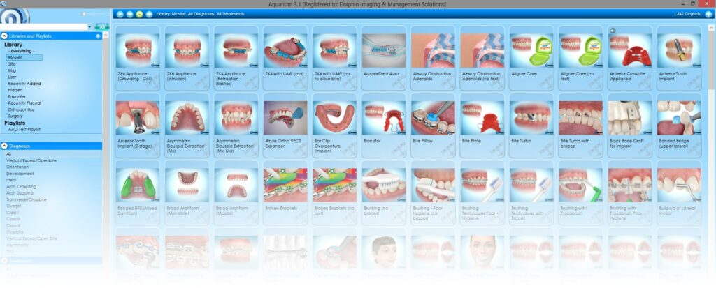
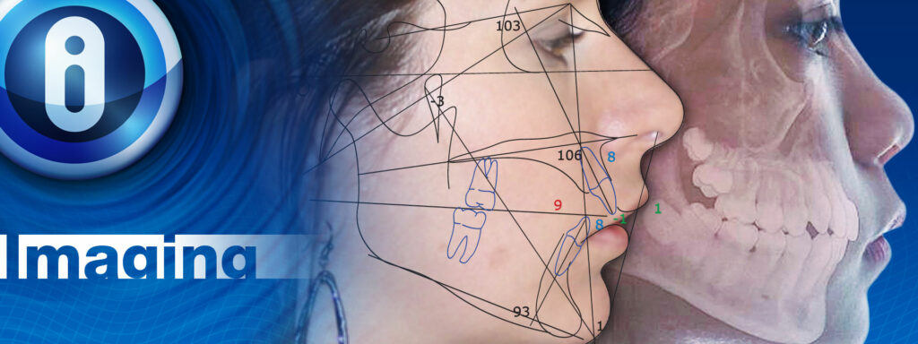
Dolphin Imaging and Management Solutions is a leading global provider in 2D/3D imaging, diagnostic, practice management and patient education software for dental specialists.


-
- Lip Bumper
-
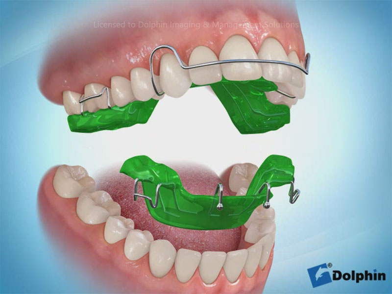
- Twin Block Appliance
-
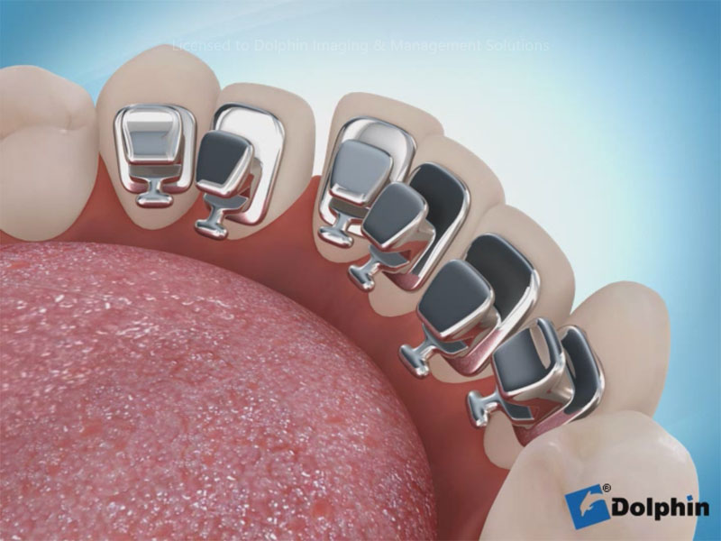
- Lingual Brackets
-
- MARA™ Appliance
-
- Spring Retainer for Retention
-
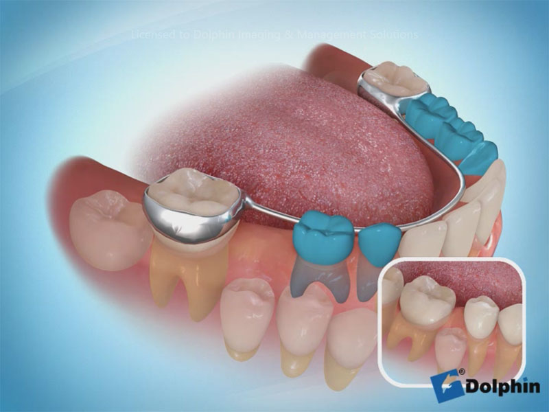
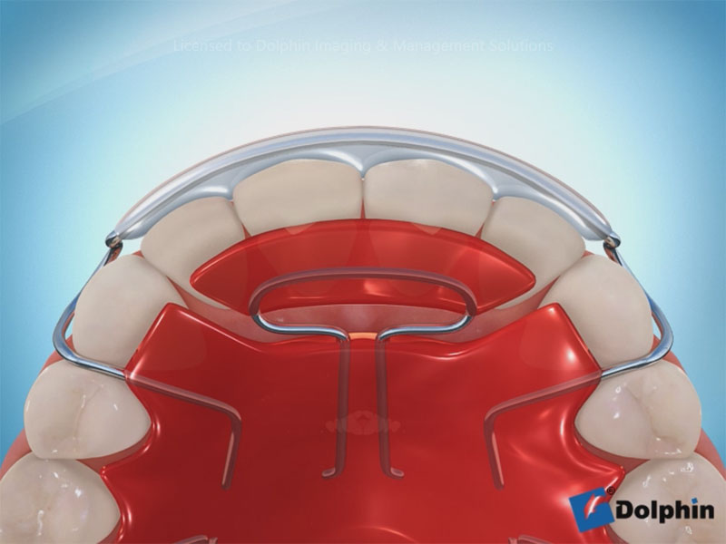
- Lower Lingual Arch
-
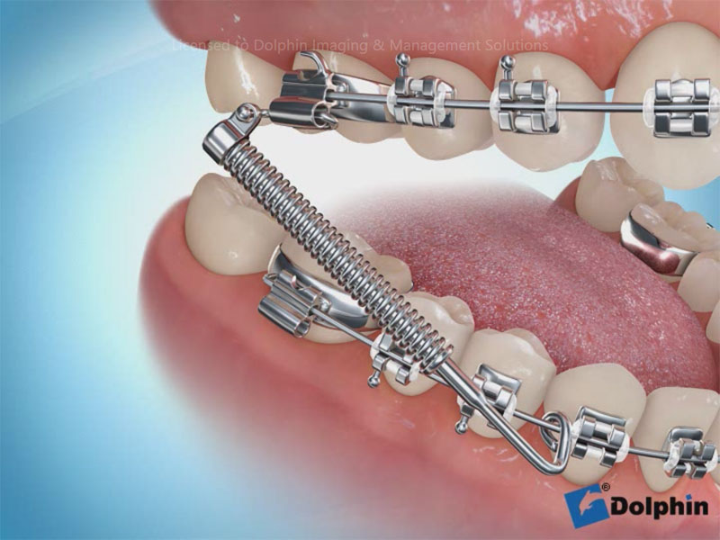
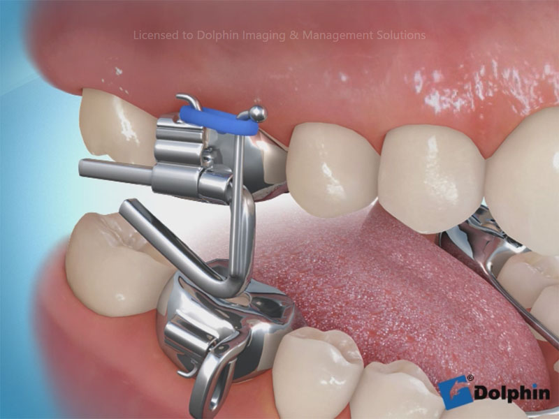
- Forsus Direct Push Rod
-
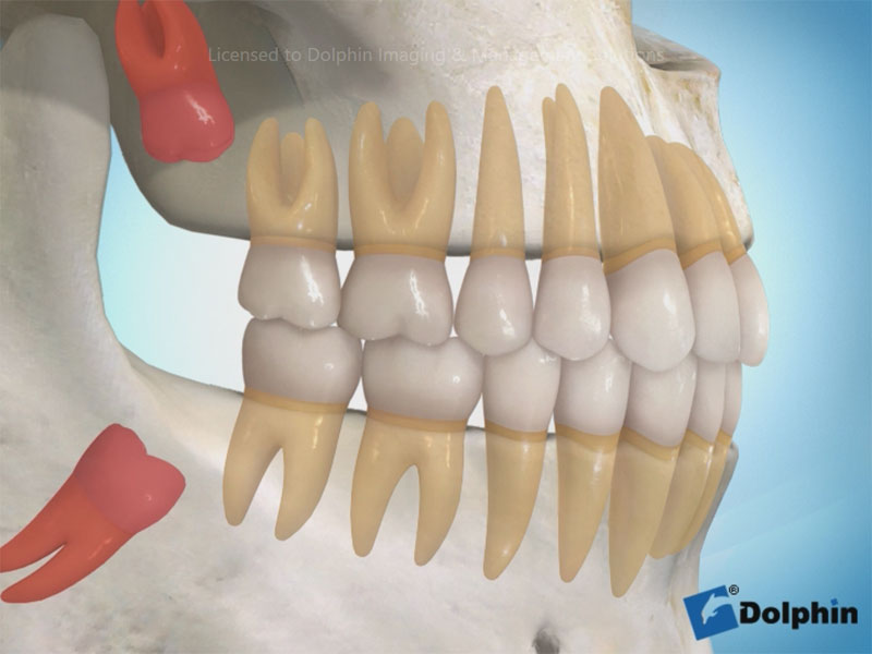
- isdom Teeth Eruption
-
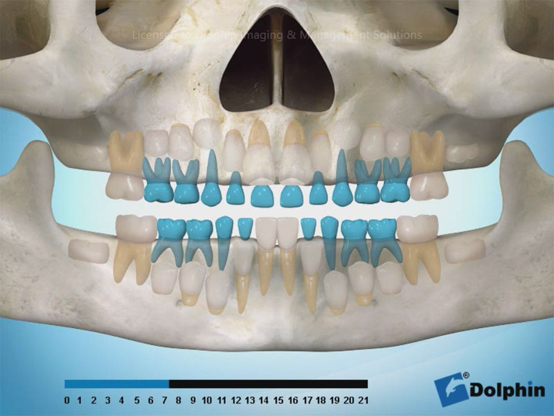
- Teeth Eruption Panoramic
-
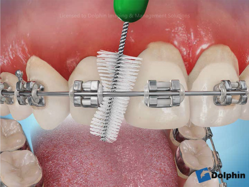
- Proxabrush Bad Hygiene
-
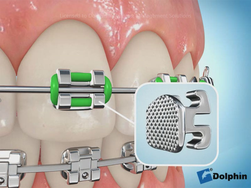
- Parts of Braces
-
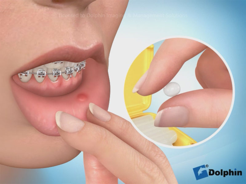
- Wax